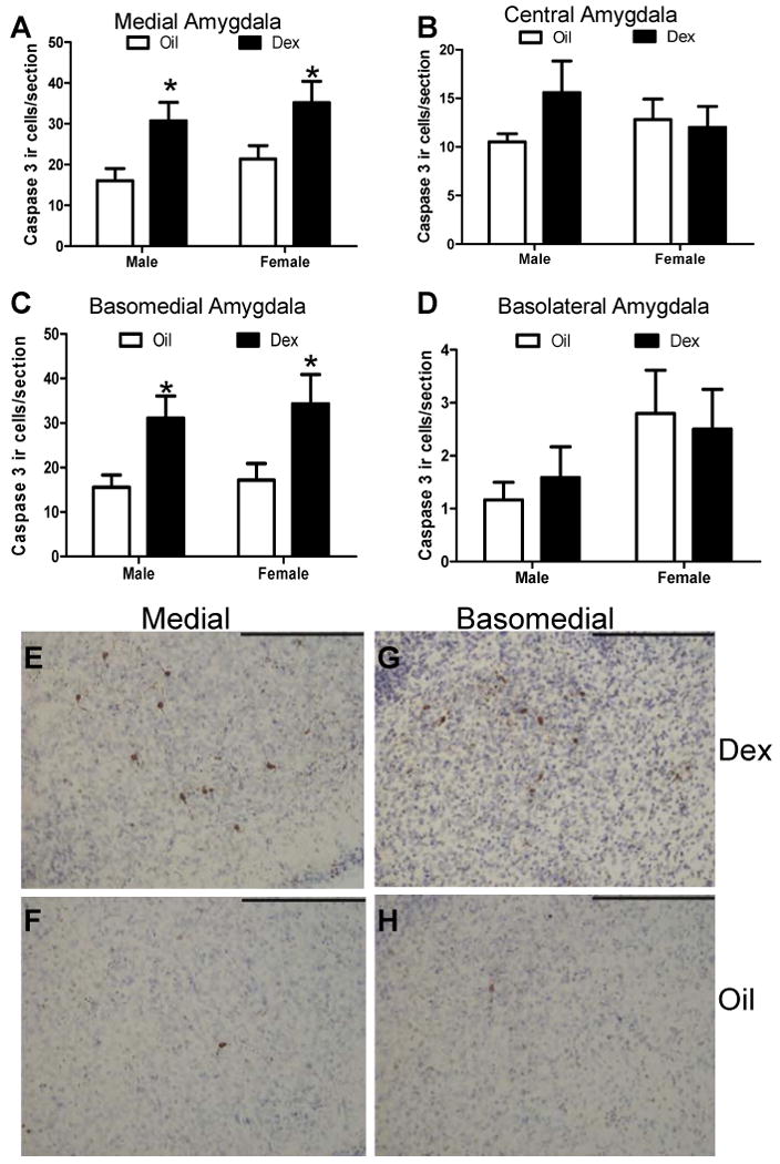
Figure 2. Cleaved caspase-3 immunoreactive cells within amygdala sub-regions following prenatal dexamethasone treatment.

The number of cleaved caspase-3 immunoreactive cells was counted in the medial (A), central (B), basomedial (C), or basolateral (D) amygdala following prenatal treatment with DEX. Representative images of cleaved caspase-3 immunoreactive cells are shown in the medial and basomedial amygdala in P0 rats following treatment with DEX (E,G) or oil vehicle (F,H) on GD18-22. Images were captured using a 20× objective in cresyl violet counterstained sections. * Post hoc comparisons indicate p<.05 compared to same sex Oil treatment. N=5 per group. Scale bar= 200μm.
