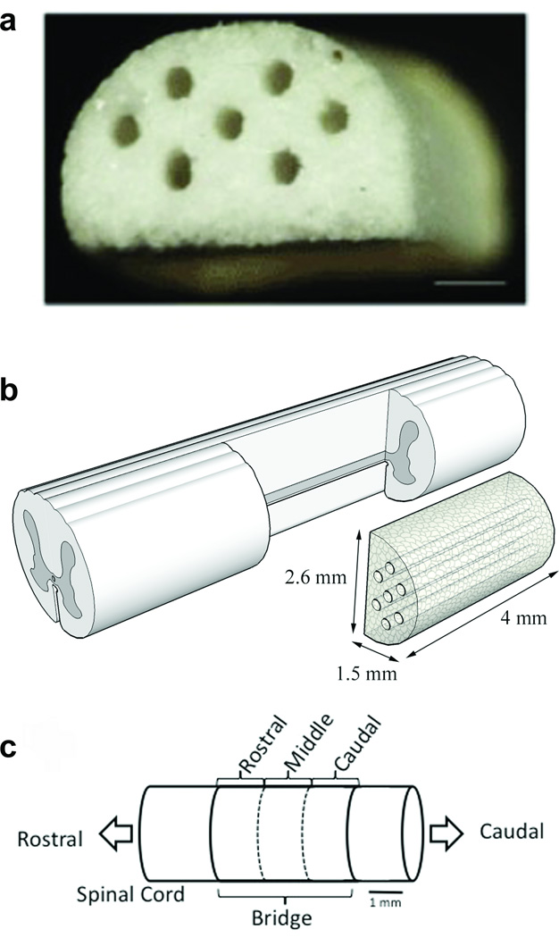
Figure 1. Multiple channel bridges for spinal cord regeneration.
(a) Photomicrograph of a multiple channel bridge showing 7 channels, each 250 µm in diameter. Scale bar is 500 µm. (b) Schematic of PLG bridge implantation in a spinal cord hemisection model. (c) Schematic of the regions in which the bridge was divided for analysis. Rostral analysis was done at 300 µm, middle at 2000 µm, and caudal at 3500 µm from the rostral edge of the bridge/tissue boundary.

