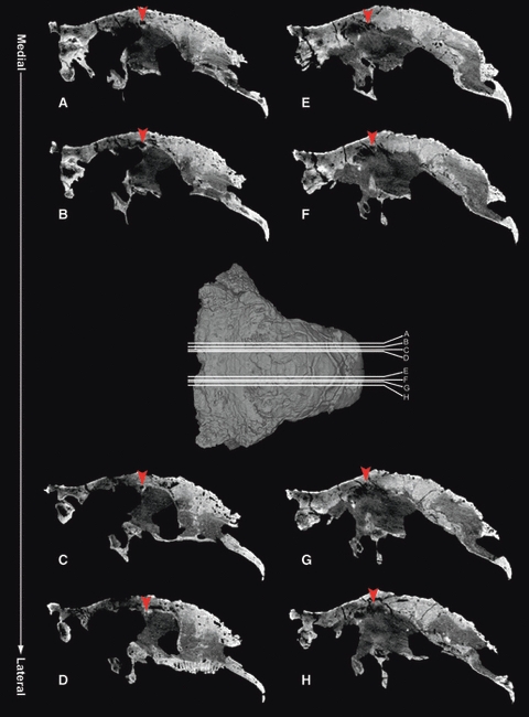
Fig. 4.

Sagittal sections of a Euoplocephalus skull (AMNH 5405) from CT data of Witmer & Ridgely (2008) show a tunnel within the frontal bone, laterally positioned and passing medially and slightly posteriorly on both sides. The most lateral sagittal section for each side is where the canal disappears into the bone. Arrowhead indicates the tunnel within the frontal, and letters A–H indicate the levels of the CT slices on the skull. Anterior is to the right in all CT slices and in the 3D model of the skull. CT data are available from the website (http://www.oucom.ohiou.edu/dbms-witmer/3D-Visualization.htm).
