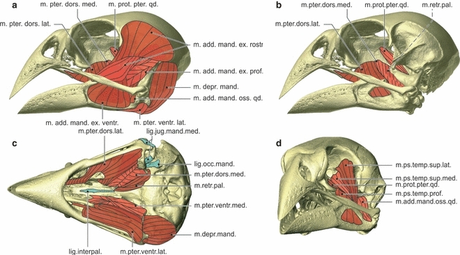
Fig. 9.

Myology Geospiza fortis. Lateral view of the skull showing the lateral muscles (a), lateral view of the skull showing the medial muscles (b), ventral view of the skull showing the ventral muscles and ligaments, with the most ventral muscles on the left side of the skull and the most dorsal muscles on the right side of the skull (c), and rostrolateral view of the skull showing the pseudotemporal muscles and the musculus adductor mandibulae ossis quadrati and the musculus protractor pterygoidei et quadrati (d). lig.interpal., ligamentum interpalatinum; lig.jug.mand.med., ligamentum jugomandibulare mediale; lig.occ.mand., ligamentum occipitomandibulare; m.add.mand.ex.rostr., musculus adductor mandibulae externus rostralis; m.add.mand.ex.ventr., musculus adductor mandibulae externus ventralis; m.add.mand.ext.prof., musculus adductor mandibulae externus profundus; m.add.mand.oss.qd., musculus adductor mandibulae ossis quadrati; m.depr.mand., musculus depressor mandibulae; m.prot.pter.qd., musculus protractor pterygoidei et quadrati; m.ps.temp.sup.lat., musculus pseudotemporalis superficialis lateralis; m.ps.temp.sup.med., musculus pseudotemporalis superficialis medialis; m.pter.dors.lat., musculus pterygoideus dorsalis lateralis; m.pter.dors.med., musculus pterygoideus dorsalis medialis; m.pter.ventr.lat., musculus pterygoideus ventralis lateralis; m.pter.ventr.med., musculus pterygoideus ventralis medialis; m.retr.pal., musculus retractor palatini.
