Fig. 3.

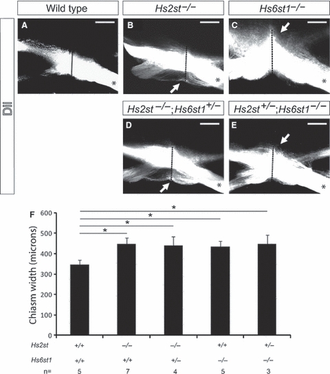
Quantitation of optic chiasm width in Hs2st;Hs6st1 compound mutants. Labelling of optic nerve following unilateral DiI injection (denoted by *) into E15.5 eyes. Labelled axons appear white in these grey-scale images and reveal the anatomy of the optic chiasm in (A) wild-type, (B) Hs2st−/− and (C) Hs6st1−/− embryos. In Hs2st−/− mutants, the optic chiasm appeared wider along the rostro-caudal midline due to the formation of an ectopic tract rostral to the chiasm (white arrow in B). In mutants lacking Hs6st1 sulphation the optic chiasm appeared wider along the rostro-caudal midline due to ectopic axon navigation along the caudal midline of the ventral diencephalon (white arrow in C). The width of the optic chiasm, taken to consist of any DiI RGC axon that could be detected using confocal microscopy, was measured along the midline (red dotted line). (F) Graph showing the width (mean ± SEM) of the optic chiasm in wild-type and compound mutant embryos. The optic chiasm was significantly wider in both the Hs6st1−/− mutant (435 ± 25 μm, n = 5) and Hs2st−/− mutant (448 ± 30 μm, n = 7) than in wild-types [345 ± 51 μm, n = 5; one way anova, multiple comparisons vs. a control group (Holm–Sidak method), P < 0.05]. The optic chiasm in Hs2st−/−;Hs6st1+/− mutants (n = 4) was 438 ± 45 μm wide, and in Hs2st+/−;Hs6st1−/− mutants (n = 3) it was 447 ± 44 μm wide. These values are not significantly different to those obtained in Hs2st−/− and Hs6st1−/− single mutants, respectively. OC, optic chiasm; ON, optic nerve; OT, optic tract. Scale bars: 200 μm.
