Fig. 2.

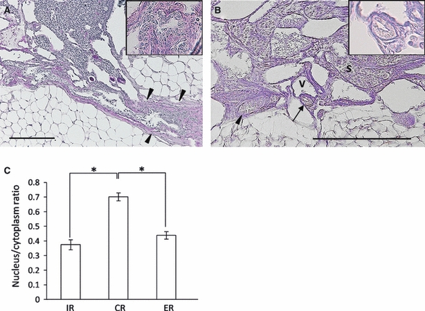
Microscopic features of the rete ovarii in B6 mice. (A) Longitudinal section of the connecting rete (CR) in a 12-month-old mouse. The CR was closely associated with the ovarian ligament (arrowheads), and its epithelial border showed an irregular line with a discontinuous basement membrane. The inset shows high magnification of the epithelium. PAS-hematoxylin stain. Bar: 200 μm. (B) Cross- (arrow) and longitudinal sections (arrowhead) of the intraovarian rete (IR) in a 5-month-old mouse. Lumen of the IR was narrow and the basement membrane was clear. The inset shows high magnification of the epithelium. PAS-hematoxylin stain. S, stroma cells; V, small vein. Bar: 200 μm. (C) Nucleus to cytoplasm area ratio in each part of the IR, CR, and ER. *P<0.0001 by the Kruskal–Wallis test, Scheffé's method, n > 30 cells in each group; value = mean ± SE.
