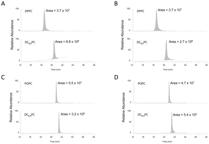
Figure 3.
Quantitation of lipids by LC-MS. Sonicated liposomes were treated with UV for 30 min at 25 °C as described in Methods section. Control samples were not irradiated. Lipids were extracted according to Bligh and Dyer protocol (see Methods section). The samples were analyzed by LC-MS. Selected reaction monitoring profiles are shown in the figure A&B, DPPC:DC8,9PC (4:1) (A) control and (B) UV-irradiated). C&D, POPC: DC8,9PC (4:1) (C) control and (D) UV-irradiated). The areas of the peaks are provided within each chromatogram.

