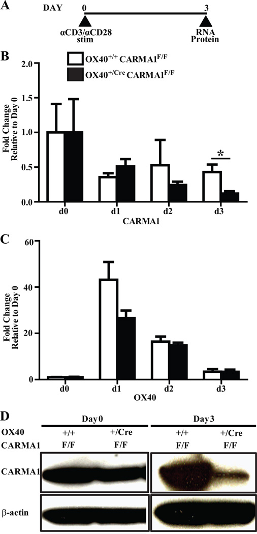
Figure 1. Deletion of CARMA1 from OX40 expressing activated CD4+ T cells.
A) CD4+ T cells isolated from mice were stimulated with anti-CD3 and anti-CD28 antibody coated beads for three days. B and C) Transcript expression of CARMA1 (B) and OX40 (C) in CD4+ T cells 0, 1, 2, and 3 days following activation, as analyzed by real-time quantitative PCR. Pooled data from 3–9 mice from 3 independent experiments are shown as mean ± SEM. *p<0.05 by t-test. D) Protein levels of CARMA1 in naïve CD4+ T cells and CD4+ T cells 3 days following activation, as analyzed by Western blot. CD4+ T cells, isolated from mouse splenocytes were activated for 3 days with anti-CD3 and anti-CD28 coated antibodies. Thirty µg total cell lysate protein was loaded for the day 0 blot and 50 µg total cell lysate protein for the day 3 blot. Following immunoblotting for CARMA1, membranes were stripped and reprobed for β-actin to ensure equal loading.

