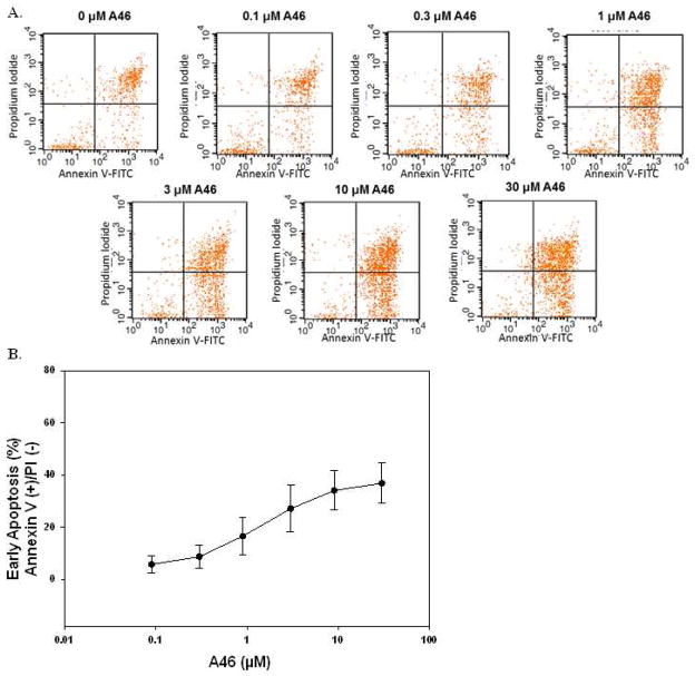
Figure 5. A46 induces apoptosis in HEL cells.
HEL cells were treated with 0, 0.1, 0.3, 1, 3, 10 and 30 μM of A46 for 48 hours, stained with annexin V-FITC and propidium iodide and then analyzed by flow cytometry to determine the level of apoptosis in the treated cells. A. Shown are representative flow cytometry profiles from one of two independent experiments. B. Quantification of the percentage of cells in early apoptosis as a function of drug treatment. Shown are the means ± S.E. of two independent experiments.

