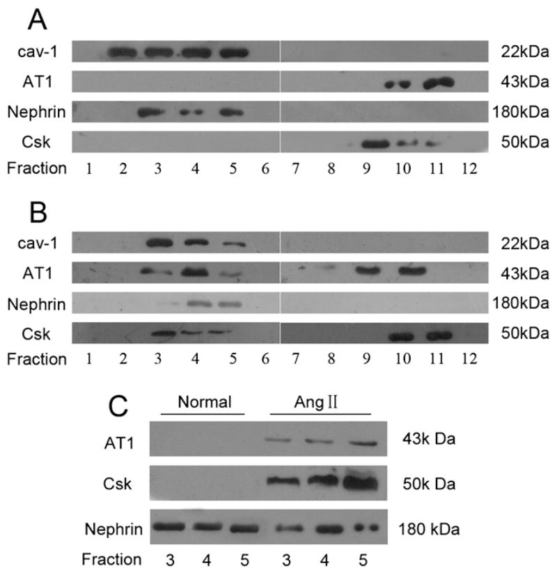
Fig. 4.
Role of caveolin-1 in Ang II-induced nephrin dephosphorylation. Lysates from normal cultured podocytes (A) and Ang II-treated podocytes for 3 h (B) were separated by sucrose gradient density centrifugation into 12 fractions as outlined in Materials and methods. (A): Fractions 2 to 5 were identified as caveolae by the presence of caveolin-1. Nephrin was also found to be present in caveolae fractions. AT1 and Csk were present in the higher density noncaveolar cytosolic fractions but not seen in the caveolae fractions. (B): After Ang II stimulation for 3 h, AT1 and Csk were found to be present both in the caveolae fractions and noncaveolar fractions. (C): Caveolin-1 was immunoprecipitated respectively from caveolae fractions (fraction 3 to 5) of normal podocytes and Ang II-treated podocytes for 3 h. The immunoprecipitates were then analyzed by Western-blotting with antibodies of AT1, Csk and nephrin. AT1: Ang II type-1 receptor; Csk: C-terminal Src kinase.

