Figure 7.

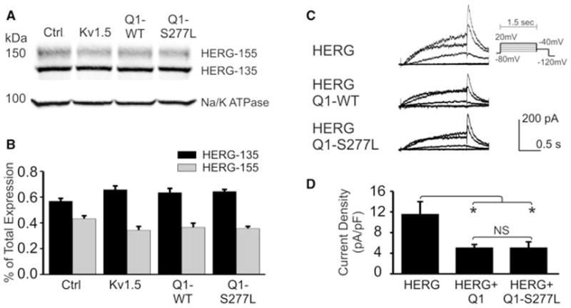
Effect of KCNQ1-WT and S277L on HERG expression and current density. (A) KCNQ1-WT (Q1-WT) and KCNQ1-S277L (Q1-S277L) were transiently transfected into HEK-293 cells that stably express HERG protein. Left lane is empty vector control, and second lane is transfected with Kv1.5. Samples were separated by 4%–15% gradient SDS-PAGE 72 hours after transfection. Western blot analysis was performed with rabbit anti-HERG and mouse anti-sodium/potassium ATPase antibodies. (B) Densitometry quantification of HERG mature (155 kDa) and immature (135 kDa) bands expressed as percent of total protein. n = 3, no statistical significance. (C) Whole cell current traces of HERG channels, from CHO cells stably expressing HERG protein and transfected with empty vector, KCNQ1-WT or S277L, patched 72 hours after transfection. Chromanol 293B was used in the bath solution to block KCNQ1 current. (D) Summary of repolarization current density measured at −40mV. n = 10, NS = no statistical significance.
