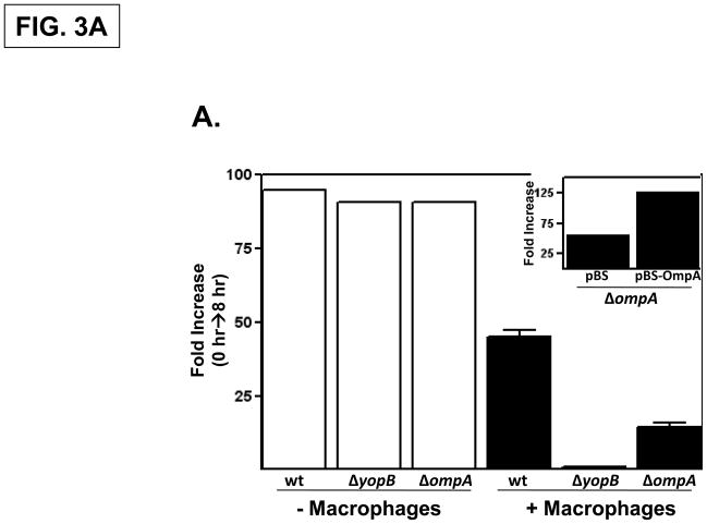
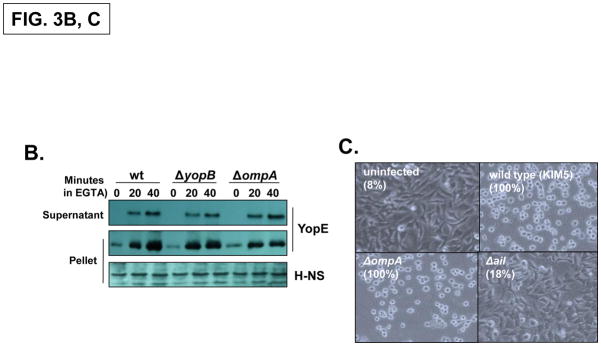
Fig. 3. The Y. pestis OmpA promotes infectivity independently of the T3SS.
(A) Empty wells (-macrophage) or wells containing RAW 264.7 cells were infected with Y. pestis KIM5, ΔyopB and ΔompA strains at a MOI of 1. Following a 30 minute attachment period, unattached bacteria were removed, and the number of cell-associated bacteria was determined by plating at the 0 hour and 8 hour timepoints. Three independent wells per strain were analyzed and the average fold-increase in the number of bacteria recovered for each strain over the 8 hour infection period is shown. (inset) The infectivity of the Y. pestis ΔompA strain was restored by a plasmid-encoded OmpA. (B) Yop secretion by the Y. pestis KIM5, ΔyopB and ΔompA strains. Strains were grown to mid-log phase and then EGTA was added to induce Yop secretion. YopE levels in both whole cells and supernatant fractions were determined by immunoblotting. H-NS levels were analyzed as a loading control. (C) HeLa cells were left uninfected or infected with wild-type Y. pestis KIM5, ΔompA or Δail strains at an MOI of 30 and photographed 2 hours after infection. Cell rounding (cytotoxicity) was quantified by counting all cells (N 200) in three different fields and the percentage is shown in parenthesis.


