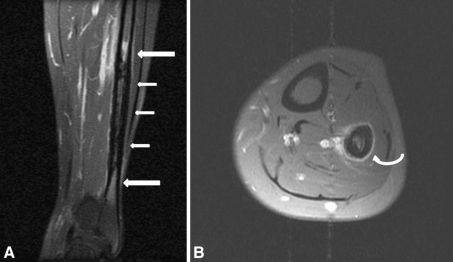
Fig. 1A–B.
MR images of the left lower leg were taken when the patient’s pain began in 1997. (A) Cortical thickening (small arrows) and bone marrow edema (large arrows) of the diaphysis of the left fibula are seen on this coronal MR image. (B) An axial gadolinium-enhanced fat-suppressed image shows periosteal reaction (curved arrow) of the left fibula.

