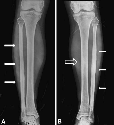
Fig. 2A–B.
Radiographs of both lower legs were obtained when the patient’s left lower leg pain worsened in February 2005, 8 years after onset. (A) Cortical thickening and increased intramedullary bone density were observed at the right fibula (arrows). There was suggestion of early subtle changes in the right midtibial diaphysis. (B) Cortical thickening, periosteal reaction, and increased intramedullary bone density were observed at the left fibula (arrows) and the left midtibial diaphysis (hollow arrow). It was most prominent at the left fibula.

