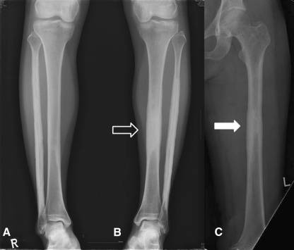
Fig. 5A–C.
Plain radiographs of the (A) right lower leg, (B) left lower leg, and (C) left femur were taken when the patient was first seen by us in 2007, 10 years after onset. It had been 2 years since her first biopsy. Hyperostosis in the left tibia had progressed with near obliteration of the intramedullary canal (hollow arrow) and newly emerged increased radiodensity was observed on the diaphysis of the left femur (arrow).

