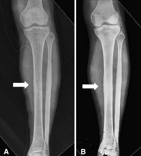
Fig. 8A–B.
Radiographs were taken immediately after the first biopsy in March 2005. (A) The tibial medullary canal was decompressed by a cortical window (arrow) created during the biopsy (see Fig. 2B for the prebiopsy radiograph). (B) A followup radiograph obtained 1 year after the first biopsy shows endosteal hyperostosis has progressed resulting in obliteration of the medullary canal (arrow).

