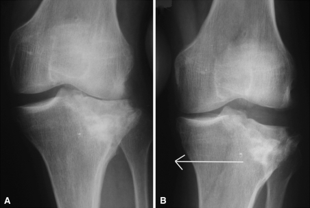
Fig. 1A–B.
(A) An AP plain radiograph of a left knee demonstrates isolated posttraumatic arthritis of the lateral compartment secondary to tibial plateau fracture malunion. A hip-knee-ankle angle of 188° was measured on full-length radiographs. (B) A varus stress view demonstrates complete correction of the valgus deformity. The arrow demonstrates the direction of stress.

