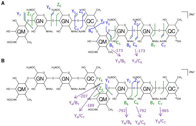
Figure 5. Multi-stage tandem mass spectra of the 18O labeled saccharide [4,2,2][0,0,0] reveals the reducing-end residue.
Tandem MS of the 18O labeled saccharide [4,2,2][0,0,0] was performed on an LTQ-Oribtrap XL mass spectrometer in positive ion mode. CID fragmentations of the precursor [M + 2Na]2+ m/z 825.2987 (A) and MS3 of the secondary product ion at m/z 835.3053 (B) are displayed on the chemical structures. Monosaccharide residues abbreviations QM (Qui4NFm), GN (GalNAcAN), and QC (QuiNAc) are shown inside the six-member rings for clarity. All fragments contain Na.

