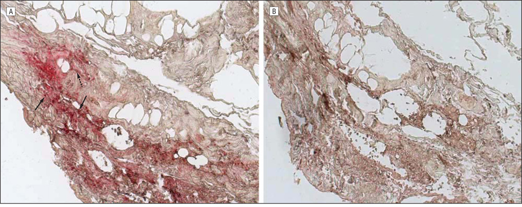
Figure 2.
Immunohistochemical analysis of the temporal artery. Note varicella zoster virus (VZV) antigen (red) in the nucleus and cytoplasm (solid arrows) and cytoplasm alone (dashed arrow) of cells in the arterial adventitia (A; original magnification ×200) not seen with normal rabbit serum (B). Sections of the temporal artery were deparaffinized and incubated with 10% normal sheep serum in phosphate-buffered saline (PBS) for 1 hour at room temperature. To prevent nonspecific binding, primary antibodies were adsorbed with normal human liver powder for 30 minutes and again for 20 hours at 4°C. Sections were then incubated with polyclonal antibodies raised against VZV open reading frame 63 protein (1:1000 dilution) or normal rabbit serum (1:1000 dilution), rinsed with PBS and incubated with a 1:300 dilution of biotinylated goat antirabbit IgG in PBS containing 5% normal sheep serum, washed 3 times in PBS, incubated with alkaline phosphatase–conjugated streptavidin (1:100 dilution), and washed 3 times with PBS. The color reaction was developed for 5 to 30 minutes with fresh fuchsin substrate system. Levamisole was added to the color reaction to block endogenous phosphatase. Uninfected and VZV-infected BSC-1 cells were used as controls (not shown).

