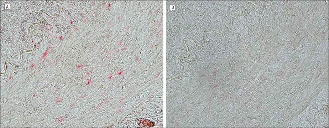
Figure 3.
Immunohistochemical analysis of the temporal artery as described in Figure 2. Note varicella zoster virus antigen scattered throughout the nucleus and cytoplasm of cells in the arterial wall (media) (A; original magnification ×400) not seen with normal rabbit serum (B).

