
Figure 10.
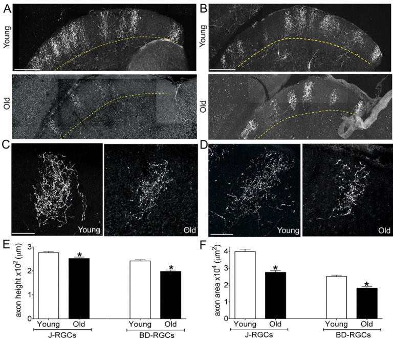
Age-related reduction in the size of RGC axonal arbors. Axonal arbors of J-RGCs (A, C) and BD-RGCs (B, D) in the superior colliculus of young adult and old mice. A, B, Low-power images of sagittal sections show that axonal arbors remain confined to the superficial half of the retinorecipient zone. C, D, High-power images of single arbors show that arbors are smaller and less dense in old than in young adult mice. E, F, Quantitative analysis showed significant age-related decreases in axon height (E) (J axons, *p = 0.002; BD axons, *p = 0.002) and axon area (F) (J axons, *p = 0.001; BD axons, *p < 0.0001). n = 24 and 38 young and old BD-RGC axons and 28 and 36 young and old J-RGC axons. Scale bars: A, B, 300 μm; C, D, 100 μm.