
Figure 8.
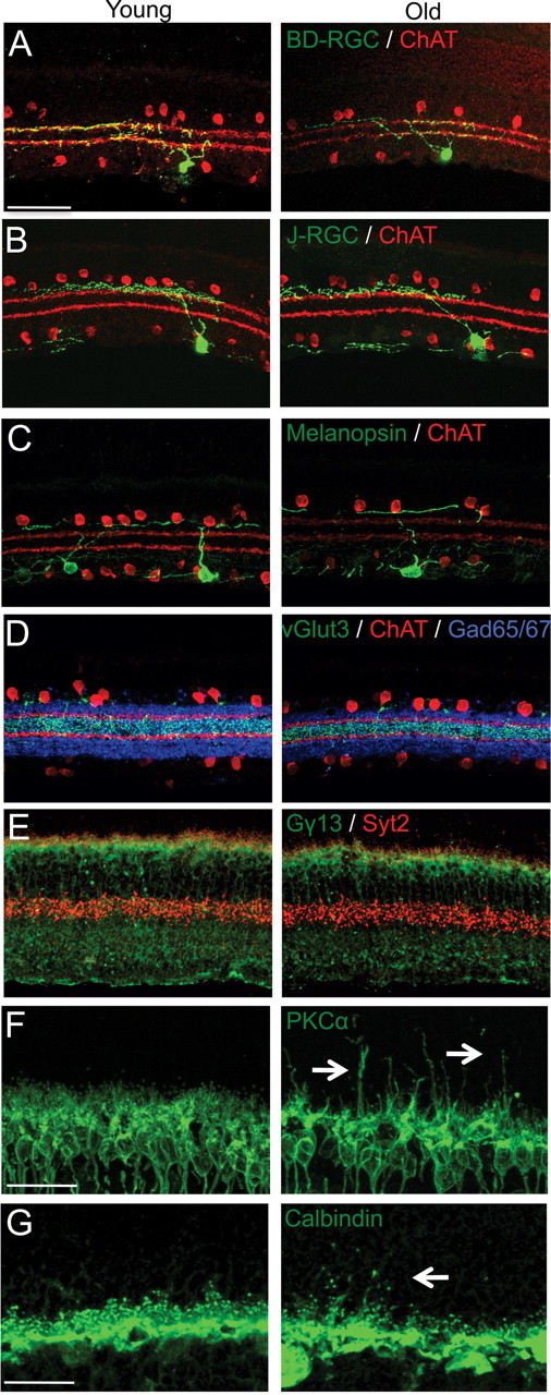
IPL lamination is intact in old mice. A–F, Sections of young adult retina and old retina were immunostained to label subsets of processes in the IPL. Scale bar, 50 μm. The outer margins of S2 and S4 are marked with anti-ChAT antibody (red) in A–D. A, GFP-positive dendrites of a BD-RGC in S2 and 4. B, GFP-positive dendrites of a J-RGC in S1. C, Melanopsin-positive RGC dendrites in S1. D, vGlut3-positive amacrine dendrites in S3 (green) and GABAergic amacrines (blue). E, Synaptotagmin-2-positive Type 2 bipolar axons in the outer IPL and (red) and Gγ13-positive ON-bipolar axons in the inner IPL (green). F, G, Sections were immunostained to label processes in the OPL. Scale bar, 25 μm. F, PKCα-positive rod bipolar cells (green) extend dendrites into the ONL in old mice (arrows). G, Calbindin-positive horizontal cell neurites (green) also send ectopic processes into the ONL (arrows) with age.