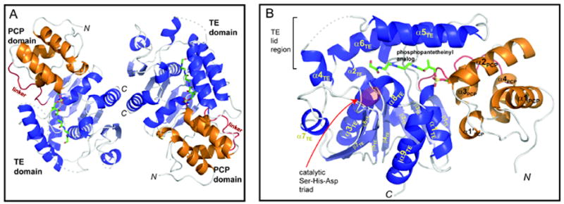
Figure 2. The overall structure of the PCP-TE conjugate.

(A), Ribbon representation of the protein dimer observed in the asymmetric unit. The PCP domain (orange) is connected to the TE domain (blue) through a short linker (red). The conjugated CoA analog is shown in licorice representation. Regions not observed in the electron density maps are indicated by dashed lines. (B), Closeup view of one of the monomer didomains. See also Figures S1, and S2.
