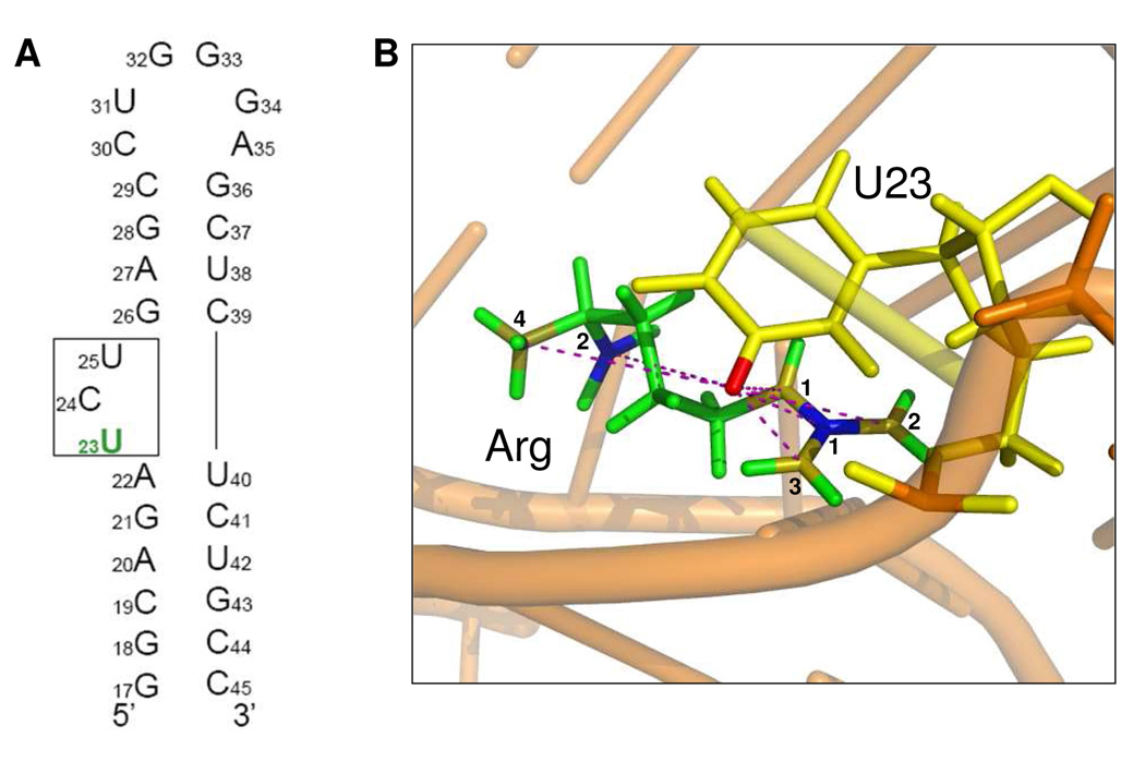
Figure 1.
a) Secondary structure of TAR RNA, indicating the position of the 5-19F base-labeled U23 nucleotide. b) Structure of the complex between TAR and a Tat-derived 37-mer peptide9. The phosphate backbone is represented by an orange ribbon and distances between TAR RNA and arginine are indicated by dash lines. Single argininamide is indicated in green with Cζ shown in blue 1, CO in blue 2, Nε in gold 1, Nη1, η2 in gold 2, 3, and NH in gold 4. U23 is indicated in yellow with the labeled 19F shown in red.

