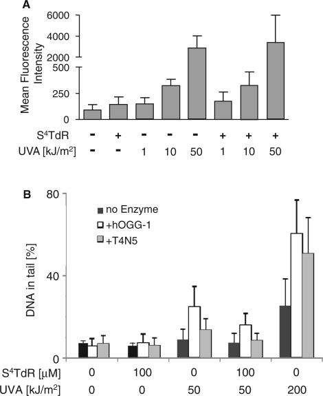
Figure 3.
(A) Reactive oxygen species. HaCaT cells were grown for 48 h in medium containing 100 μM S4TdR to replace ∼0.1% of DNA thymidine. PBS-washed cells were incubated with the ROS-sensitive probe CM-H2DCFDA, irradiated with UVA as indicated and ROS-induced fluorescence measured by flow cytometry. Data are presented as the mean fluorescence intensity ± standard deviation of three experiments. (B) DNA single-strand breaks/guanine oxidation/cyclobutane pyrimidine dimer (CPD) formation. HaCaT cells grown in the presence or absence of 100 µM S4TdR were washed and irradiated with 50 or 200 kJ/m2 UVA as indicated. Direct DNA single-strand breakage was analysed by the alkaline comet assay (No enzyme, black bars). The presence of DNA 8-oxoguanine was revealed by digestion with hOGG-1 (+hOGG-1, open bars) and CPDs by digestion with T4N5 (+T4N5, grey bars) as indicated. DNA damage is expressed as percentage of DNA in the comet tail.

