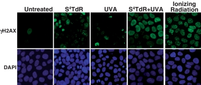
Figure 4.
γ-H2AX staining. HaCaT cells grown in the presence of 100 µM S4TdR for 48 h were irradiated with 10 kJ/m2 UVA as shown. Control cells grown in the absence of S4TdR were treated with 10 Gy of gamma irradiation. In each case, γ-H2AX was visualized 4 h later. Note the pan-nuclear γ-H2AX staining induced by S4TdR/UVA treatment which differs from the discrete focal staining following gamma irradiation. DNA was counterstained with 4′,6-diamidino-2-phenylindole (DAPI).

