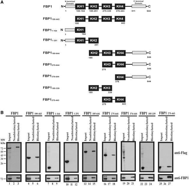
Figure 3.
Identification of interaction domains in FBP1 for EV71 5′-UTR. (A) Schematic diagram of various truncated forms of FBP1. The black boxes indicate KH domains. The N-terminal domain and the C-terminal domain are as indicated. Fused flag tags at the N-terminals of various truncated forms of FBP1 were applied in a pull-down assay. The numbers of the truncated form FBP1 indicate first and last amino acids. (B) Map interaction regions in FBP1 for EV71 5′-UTR. Wild-type FBP1 (lane 1) or various truncated forms of FBP1 (lanes 4, 7, 10, 13, 16, 19, 22 and 25) expression plasmid were transfected into RD cells. Cell extracts from various transfected forms of FBP1 were collected and incubated with biotinylated EV71 5′-UTR RNA probe (lanes 3, 6, 9, 12, 15, 18, 21, 24 and 27) or non-biotinylated RNA probe (lanes 2, 5, 8, 11, 14, 17, 20, 23 and 26). Western blot using anti-Flag and anti-FBP1 antibodies was applied to examine protein expression. The RNA–protein complex with beads was resolved for SDS–PAGE (12%).

