Figure 5.
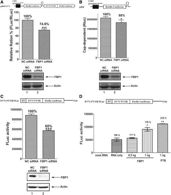
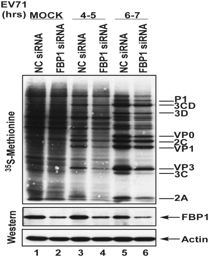
Viral IRES activity and viral protein synthesis were positively regulated by FBP1. (A) Schematic diagram of dicistronic reporter plasmids pRHF-EV71. Plasmid expresses dicistronic mRNA, consisting of cytomegalovirus (CMV) promoter, the first cistron RLuc gene, the EV71-5′-UTR and the second cistron FLuc. A hairpin (H) is inserted downstream of the first cistron to prevent ribosome read-through. RD cells were transfected siRNA against FBP1. After 3 days, dicistronic construct pRHF-EV71 and FBP1 siRNA were co-transfected into RD cells. After 2 days, the RLuc and FLuc activity in cell lysates were analyzed. The bars in the histogram represent FLuc/RLuc activity percentages. Experiments were performed in triplicate to obtain the bar graph. Western blotting was utilized analyze the expression levels of FBP1 and actin. (B) Schematic diagram of cap-dependent reporter pRH. RD cells were transfected FBP1 siRNA firstly. After 3 days, cap-dependent reporter construct pRH and FBP1 siRNA were co-transfected into RD cells. After 2 days, the RLuc activity in cell lysates were analyzed. Western blotting was utilized analyze the expression levels of FBP1 and actin. (C) Schematic diagram of monocistronic reporter EV71-5′-UTR-FLuc. Monocistronic mRNA containing EV71 IRES and FLuc was transfected to cells pre-treated with FBP1 siRNA or NC siRNA. At 6 h post-transfection, the RD cell lysate was assayed for FLuc activity. Cells were harvested, lysed and western blotted for FBP1 and actin. (D) In vitro IRES activity assay was performed contained monocistronic reporter RNA (EV71-5′-UTR-FLuc), different amounts recombinant FBP1 proteins or PTB, HeLa cells translation extracts and 20% RRL. The mixtures were incubated and measured FLuc activity. (E) Viral proteins synthesis in FBP1 knockdown cells. RD cells transfected with of NC siRNA or FBP1 siRNA were challenged with EV71 and subjected to a pulse-labeling assay. Protein synthesis in mock-infected (lanes 1 and 2), and EV71-infected cells were examined by 35S-methionine pulse-labeling at various times (lanes 3–6). Western blot analysis of FBP1 protein knockdown efficiency was performed (lower panels). (*P < 0.05, **P < 0.01 and ***P < 0.001, Student's two-tailed unpaired t-test).


