Figure 3.
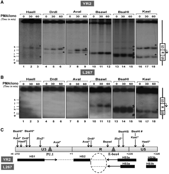
PMA/ionomycin-mediated disruption of the chromatin organization within the BLV promoter region. Nuclei from YR2 (A) or L267 (B) cells mock treated or treated with PMA/ionomycin (for 30 or 60 min) were digested by restriction enzymes (HaeII, DrdI, AvaI, BsaWI, BsaHI and KasI) and examined by indirect end-labeling following BamH1 digestion in vitro. When several restriction sites for the same enzyme were found within the region under study, symbols were used (open circle, asterisks, dollar and hash) to differentiate them. The Figure shows one representative experiment from three independent IEL assays. (C) Schematic representation of HS positions in the YR2 and L267 5′-LTR (solid bars) and of the regions which became accessible following PMA/ionomycin stimulation (indicated by arrows). Positions of restriction sites of interest and of the PU.1 an E-box 4-binding sites are indicated.

