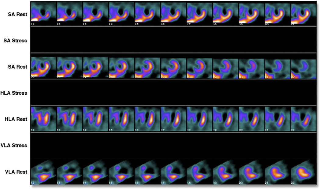
Figure 3. Example of measurable infarct size.
Rest only images of a 47-year-old-man who presented 21 hours after onset of chest pain with an anterior wall STEMI treated with direct PCI. Initial c-TnT was 4.55 ng/mL. C-TnT at day 3 was < 0.01 ng/mL. SPECT-MPI showed a large anterior, apical, antero-lateral and antero-septal perfusion defect. Infarct size was quantitated at 55% of the LV. LVEF was 32%.

