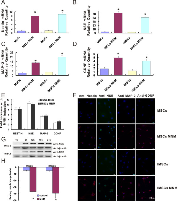
Figure 5.
IMSCs exhibit the same neuronal differentiation capacity as primary MSCs in vitro. A-D: Transcriptional expression of Nestin, NSE, MAP-2, GDNF during neuronal differentiation of MSCs and IMSCs. The transcriptional expression of genes was determined by measuring their mRNA levels with real-time PCR. Treatment of primary MSCs and IMSCs with MNM resulted in a dramatic increase in the mRNA levels (*, P < 0.001 by Student's t test). Real-time PCR results were confirmed in at least three batches of independent experiments with β-actin normalization. E: Fold increase of Nestin, NSE, MAP-2, and GDNF mRNA expression levels after induced by MNM. The mRNA expression levels of neural markers in both MSCs and IMSCs increased about 8, 10, 5 and 3 fold increases after treated with MNM, respectively (P > 0.05 by Student's t test). F: Immunofluorescence staining of Nestin, NSE, MAP-2, and GDNF during neuronal differentiation of MSCs and IMSCs (Scale bar = 20 μm). G: Expression of NSE during a 24 hr span including 0, 6, 12, 18, and 24 hr time-point of MNM induction in both primary MSCs and IMSCs were analyzed by western blotting. Equal loading of the samples was confirmed by comparable levels of β-actin detected in each lane. H: Resting membrane potential was measured by using whole cell patch clamp. The values represented Means ± S.E.M. (*, p < 0.001, MSCs vs. MSCs + MNM; &, p < 0.001, IMSCs vs. IMSCs + MNM; p > 0.05, MSCs + MNM vs. IMSCs + MNM, by Student's t test).

