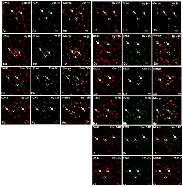
Figure 2.
P2X4 immunoexpression in P0 rats subjected to hypoxia (Hy) at different time points. P0 rats subjected to Hy and sacrificed at 4 (Hy4 h), 24 (Hy24 h), 72 (Hy72 h), 7D (Hy7 d) and 14D (Hy14 d). Note P2X4 immunofluorescence in amoeboid microglial cells overlapping with OX42 fluorescence is noticeably enhanced in the hypoxic rats Hy4 h (A/Ba-c), Hy24 h (C/Da-c), Hy72 h (E/Fa-c), and Hy7 d (H/Ia-c) when compared with the matching controls (Con4 h, Con24 h, Con72 h, Con7 d and Con14d). Scale bar = 20 μm.

