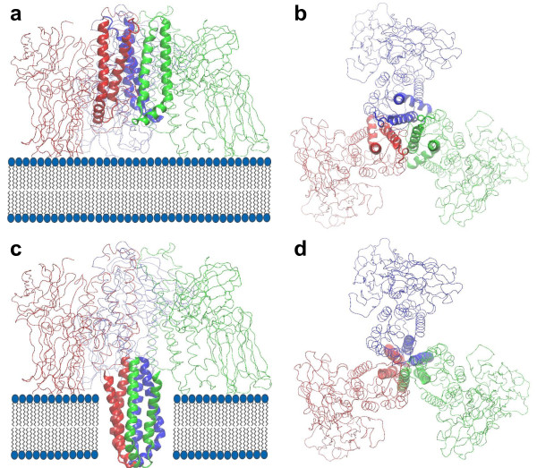
Figure 8.
Pore formation of the pre-pore Cry4Aa trimer by insertion of three α4-α5 hairpins into a membrane bilayer. (a) side view, and (b) top view before insertion, (c) side view, and (d) top view after the insertion of the three hairpins with 90° hairpin rotation to form a barrel that has a channel at the center. The three hairpins are rendered as ribbons and colored blue for M1, red for M2, and green for M3. Other parts of the protein are drawn as lines.

