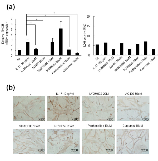
Figure 4.

IL-17-mediated RAGE induction in RA-FLS involves PI3 kinase, STAT3, NF-κB, and AP-1. (a) RA-FLS were pretreated with 20 μM LY294002, 50 μM AG490, 10 μM SB203580, 20 μM PD98059, 10 μM parthenolide, or 10 μM curcumin for 30 minutes, and then 10 ng/ml IL-17 was added for 12 h. RAGE mRNA was analyzed by real-time PCR. RA-FLS were cultured as in Figure 4a. The lactate dehydrogenase (LDH) concentrations in the culture supernatants were determined using an activity assay kit. (b) FLS were treated with same method as (a). RAGE expression in the FLS was determined using a RAGE-specific antibody. The brown color shows the RAGE. Values are the mean ± SEM of triplicate cultures. *P < 0.05 compared to inhibitor-treated cells.
