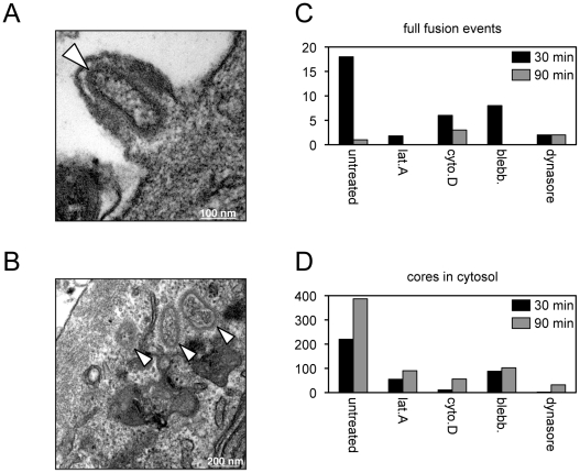
Figure 4. Effects of inhibitors on entry determined by transmission electron microscopy.
Purified MVs (350 PFU per cell) were spinoculated onto inhibitor-treated HeLa monolayers at 4°C for 60 min. Virus-bound cells were then incubated for either 30 or 90 min at 37°C in the presence or absence of the indicated inhibitor, fixed and processed for transmission electron microscopy. Representative images from untreated cells at 30 min showing full fusion of virion and plasma membranes resulting in pore formation (A) and cores in the cytosol (B). White arrowheads point to cores; scale bars indicate magnification. For each infection, a total of 90 randomly-selected cell sections were visualized and the number of plasma membrane full fusion events (C) and viral cores in the cytosol (D) were determined at 30 and 90 min.

