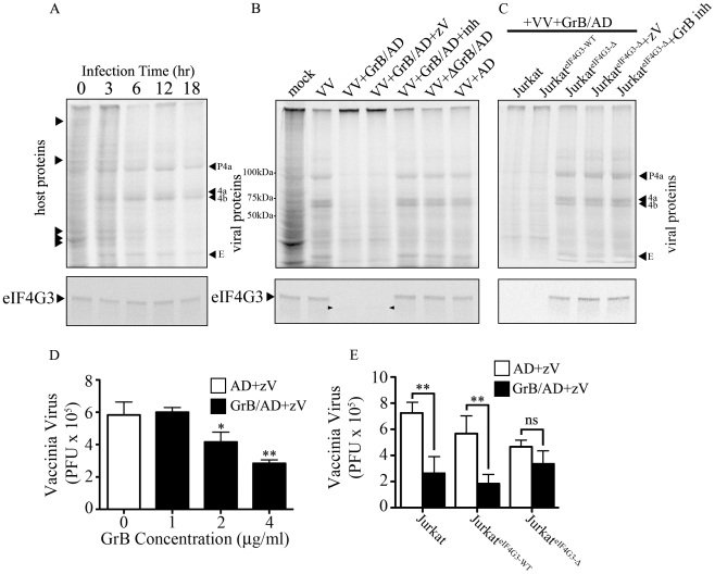
Figure 6. Inhibition of VV production by GrB is mediated by cleavage of eIF4G3.
(A) Jurkat cells were infected with VV for 0 to 18 hr. Metabolic labeling was assessed by MetS35 incorporation and viral protein synthesis was monitored by autoradiography analysis. Arrowheads on the left hand side of the radiogram indicate host proteins and arrowheads on the right hand side of the radiogram indicate VV proteins. Late VV proteins P4a, 4a, 4b and early VV protein E are shown. Western blot analysis of eIF4G3 from each time point is shown below the radiogram. (B) Jurkat cells were infected for 12 hr with no virus (mock) or with VV. Infected cells were co-treated with AD, GrB/AD, GrB/AD+zVAD−fmk (zV), GrB/AD+GrB inhibitor (inh) and heat inactivated GrB+untreated AD (ΔGrB/AD). Western blot analysis of eIF4G3 from each experimental condition is shown below the radiogram. Arrowheads in the Western blot image point to eIF4G3 degradation product; (n = 4 of 4 independent experiments). (C) Jurkat cells, Jurkat cells transformed with wild-type eIF4G3 (JurkateIF4G3-WT) and Jurkat cells transformed with GrB-resistant mutant (JurkateIF4G3-Δ) were infected with VV for 12 hr (D) The effect of GrB concentration (1–4 µg/ml) on VV replication was tested. AD and zV concentrations were the same in all treatments; Statistical significance: p<0.05 (*) and p<0.01 (**); where * compares 2 and 4 µg/ml GrB vs. 0 µg/ml GrB; (n = 3 of 3 independent experiments). (E) VV replication in untreated Jurkat cells, JurkateIF4G3-WT, and JurkateIF4G3Δ, was measured by plaque assay. Statistical significance: p<0.01 (**); (n = 3 of 3 independent experiments).

