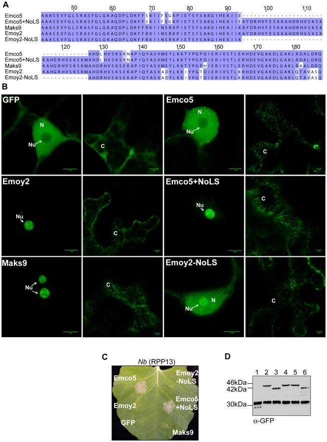
Figure 5. Nucleolar targeting signal of ATR13. A.
An alignment of various ATR13 chimeras showing the naturally occurring insertion present in Maks9 and Emoy2 alleles of ATR13, the insertion added to the Emco5 allele, and the deletion from the Emoy2 allele. B. Localization of GFP-fused ATR13 chimeras expressed transiently in N. benthamiana. The left panels are focused on nuclei (N) and nucleoli (Nu), whereas panels to the right are images of associated cytoplasm (C). Scale bars are 5 um. C. Expression of these constructs in N. benthamiana containing RPP13Nd showing intact recognition patterns despite altered localization. D. Western blot of various ATR13 alleles and chimeras probed with α-GFP showing comparable expression levels in N. benthamiana. Lanes are labeled as following: 1. 35S-GFP, 2. 35S-Emoy2:GFP, 3. 35S-Emco5:GFP, 4. 35S-Maks9:GFP, 5. 35S-Emco5+ NoLS:GFP, 6. 35S-Emoy2-NoLS:GFP.

