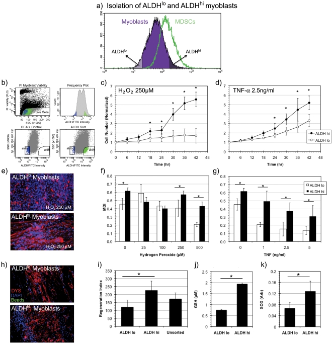
Figure 2. Isolation and stress resistance capacity of ALDHhi myoblasts.
(a, b) ALDHhi murine myoblasts were isolated via fluorescence activated cell sorting (FACS) from a heterogeneous population of skeletal muscle derived cells using DEAB, a potent inhibitor of ALDH, as a gating control. (c, d) A significantly increased rate of proliferation of ALDHhi myoblasts compared to ALDHlo myoblasts was observed in conditions of oxidative stress (H2O2, 250 µM) (* indicates p<0.05) and inflammatory stress conditions (TNF-α, 2.5 ng/ml) (n = 9). (e) ALDHlo and ALDHhi myoblasts underwent myogenic differentiation by fusing into MHC+ myotubes (red) under oxidative stress conditions (H2O2, 250 µM). Nuclei were stained with DAPI (blue). (f) Significantly increased myogenic differentiation indices (MDI) were observed in ALDHhi cells at several concentrations of oxidative stress (0, 250 and 500 µM) when compared to ALDHlo (n = 3). (g) Similarly increased MDIs of ALDHhi cells were observed under all inflammatory stress conditions when compared to ALDHlo myoblasts. (h) An increased number and density of dystrophin positive myofibers (stained in red) were observed in mdx mice injected intramuscularly with ALDHhi cells compared to those injected with ALDHlo myoblasts. Nuclei are DAPI (blue) stained. Green FluoSphere beads are observed to localize at the area of initial injection. (i) A significantly increased regeneration index was observed in ALDHhi transplanted mice compared to those injected with ALDHlo and unsorted cells. (n = 3) (j, k) Measurements of intracellular antioxidants glutathione (GSH) and superoxide dismutase (SOD) levels demonstrated statistically significant elevation of both antioxidants in ALDHhi myoblasts compared to ALDHlo myoblasts (n = 3).

