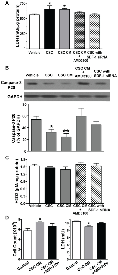
Figure 9. CSC-derived SDF-1 in the attenuation of cellular injury.
A, Remaining LDH levels in cardiac tissue were determined after I/R. B, Western blot analysis indicated cleaved caspase-3 levels after I/R. Shown is representative immunoblots in each groups (one lane/group). Bar graph represents relative levels of Caspase-3 P20 (% of GAPDH). C, Myocardial hydrogen peroxide (H2O2) production was analyzed after I/R. A–C: Mean ± SEM, N = 4–5/group, *p<0.05, **p<0.01 vs. Vehicle. D, Cardiomyocyte (H9c2) viability and LDH levels in supernatant were determined after 24-hr of hypoxia in groups of vehicle and CSC CM with or without AMD3100. Mean ± SEM, N = 3, *p<0.05 vs. vehicle.

