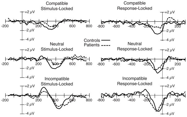
Figure 3.
Stimulus-locked (left) and response-locked (right) grand average ERP difference waveforms (contralateral-minus-ipsilateral) for the compatible, neutral, and incompatible stimulus categories, collapsed across the C3 and C4 electrode sites, with patient and control waveforms overlaid. Stimulus-locked waveforms were time locked to the onset of the target stimulus.

