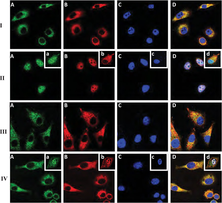
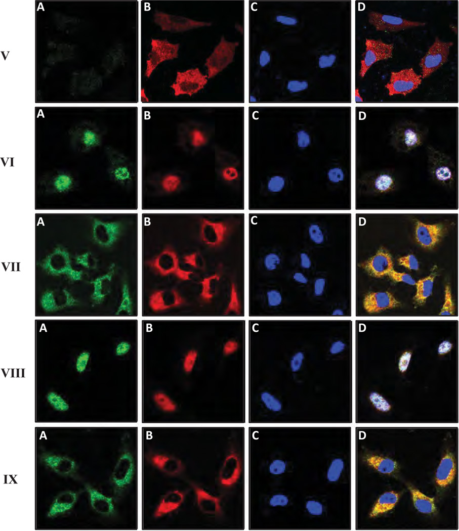
Figure 4.
Immunofluorescence staining of MyD88 and NFκB-p65 in MDA-MB-231 cells: MDA-MB-231 cells treated with various agents were fixed by 2% formaldehyde. Subsequently, cells were rendered permeable by ethanol treatment and stained with FITC-labeled anti-MyD88 antibody (green color) (A or a), Texas Red-labeled NFκB-p65 antibody (red color) (B or b) or DAPI (blue color) (C or c) and an overlay image (D or d) of (A, a) (B, b) and (C, c) as described in the Materials and Methods.
MDA-MB-231 cells were treated with no HA (I); or with LMW-HA (II) [or with HA fragments (2–3 disaccharides) (inset, a–d)]; or with HMW-HA (III); or with anti-CD44 antibody followed by LMW-HA (IV) [or with normal IgG plus LMW-HA, inset, a–d)]; or transfected with MyD88 siRNA plus LMW-HA (V); or with scrambled siRNA plus LMW-HA (VI); or with AFAP-110 siRNA plus LMW-HA (VII); or with scrambled siRNA plus LMW-HA (VIII); or with cytochalasin D plus LMW-HA (IX).


