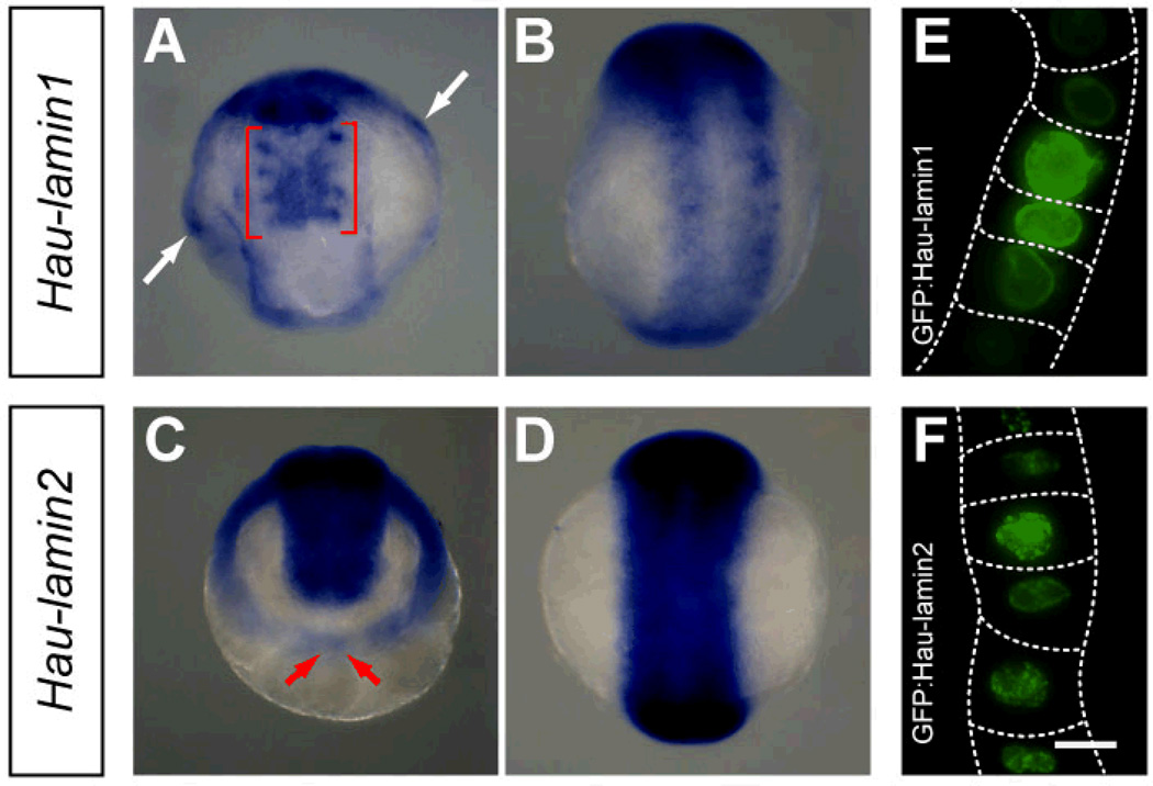
Fig. 7.
Characterization of Helobdella lamin gene expression. A Hau-lamin1 is first detected in late stage 8 (anterior ventral view); it is expressed in the nephridium-like mesodermal structures in the four rostral segments (red brackets) and in cells of the provisional integument (white arrows). B In stage 9 (ventral view), Hau-lamin1 is expressed in the developing definitive epithelium, which also expresses Hau-cif1 and Hau-cif9. C Hau-lamin2 is expressed in the germinal bands in stage 8 (animal pole view of an embryo at mid stage 8 is shown in C); Hau-lamin2 expression is weaker in the more posterior region of germinal bands (red arrows). D In stage 9 (ventral view), Hau-lamin2 is strongly expressed throughout the germinal plate. E, F Fluorescence images of blast cells expressing GFP:lamin fusion protein; the GFP fluorescence exhibited strong nuclear localization for both GFP:Hau-lamin1 (E) and GFP:Hau-lamin2 (F). Dotted line marks approximate outline of blast cells expressing GFP-lamin fusion protein. Scale bar: 120 µm (A–D); 10 µm (E, F).

