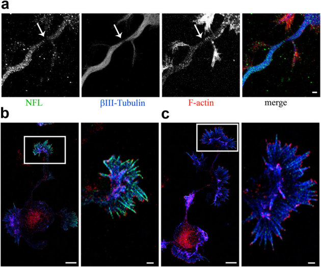
Figure 4. The laser damage induces a partial dissection of the three cytoskeletal elements.

BDNF increases actin filopodia at steady state. (a) Immunostaining of neurofilament subunit L (green), βIII-tubulin (blue) and F-actin (red) in the axon of a hippocampal neuron at 3DIV immediately after laser injury. Bar is 2 µm. (b) Immunostaining of actin (green), F-actin (blue) and Abi-1 (red) in a hippocampal neuron at 3DIV and (c) in a hippocampal neuron at 3DIV treated with 100 ng/ml BDNF for 30 minutes. The right panels, of b and c, show magnifications of the growth cones (white square box in the left panels). Bars are 10 µm in the left panels, and 2 µm in the right panels.
