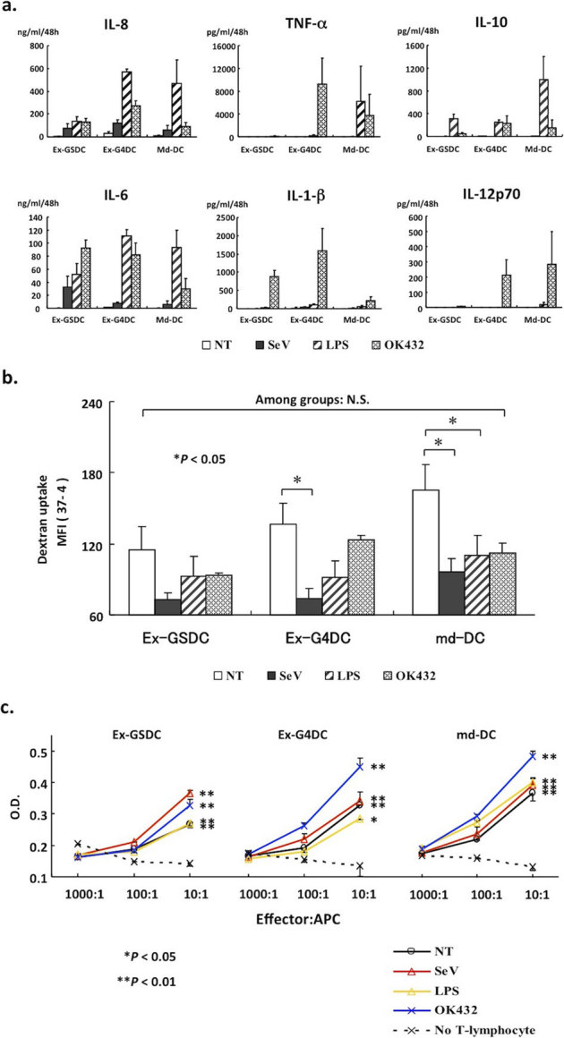
Figure 3. Assessment of functions that are typically seen in DCs.

(a) Expression of typical human inflammatory cytokines/chemokines of monocyte-derived DCs and expanded DCs in response to various stimuli. These six panels were assessed by the Cytometric Bead Array (CBA) system and contain data from three independent experiments. (b) FITC-dextran uptake assay assessing endo-/phagocytotic activity, a typical feature of antigen-presenting cells like DCs. DCs were stimulated by each stimulus and then exposed to 1 mg/ml of FITC–dextran for 30 min at 4 or 37 degrees Celsius. The uptake was expressed MFI between cell samples incubated at 37 and 4 degrees Celsius. This experiment was performed three times. (c) A graph showing MLR activity for allo-antigen by each immature DC or activated DC by SeV/dF, LPS, or OK-432. This experiment was performed three times.
