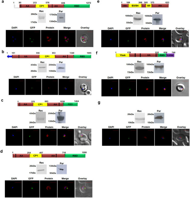
Figure 2. Targeting of P. falciparum aaRSs and of DTD.

Localization of (a) Pf-Ed-IRS1 (b) Pf-Ed-IRS2 (c) Pf-Ed-LRS (d) Pf-Ed-VRS (e) Pf-Ed-FRS (f) Pf-Ed-PRS (g) Pf-DTD. In all cases, upper panels show name of P. falciparum aminoacyl-tRNA synthetase, and their domain/ subdomain features. Middle panels show P. falciparum aminoacyl-tRNA synthetase expression in parasites (Par) and detection of recombinant P. falciparum aminoacyl- tRNA synthetase domains (Rec) by western blot analysis. Lower panels display their cellular localizations. Editing domains are colored yellow, aminoacylation domain (AA) is in red; RNA binding domain (RBD) is in green; ProRS specific C-terminal domain is in purple and un-annotated domains are in white. Blue arrow represents apicoplast targeting sequences predicted by PATS. Conserved motifs are highlighted by black strips. The parasite line used was GFP-tagged (strain D10 ACPleader-GFP) where apicoplast fluorescence is in green. DAPI staining is in blue while aminoacyl-tRNA synthetases are stained with Alexa594 (red).
