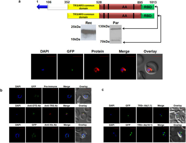
Figure 3. Expression and localization of Pf-Ed-TRS.

(a) Upper panel shows P. falciparum aminoacyl-tRNA synthetase name, and domain/ subdomain features. Middle panel shows P. falciparum aminoacyl-tRNA synthetase expression in parasites (Par) and detection of recombinant P. falciparum aminoacyl- tRNA synthetase domains (Rec) by western blot analysis. Lower panel displays their cellular localizations. Editing domains are colored yellow, aminoacylation domain (AA) is in red; RNA binding domain (RBD) is in green. Blue arrow represents apicoplast targeting sequences predicted by PATS. Conserved motifs are highlighted in black strips. (b) Upper and lower panels show confocal IFA with pre-immune sera and with anti-histidine antibodies, whereas the middle panels depict cytoplasmic staining of Pf-DTD. (c) Upper and lower panels show results of competitive confocal IFAs with rabbit anti-Ed-TRS antibodies which were pre-incubated with Ed-TRS in molar ratios of 1∶1 and 10∶1 respectively.
