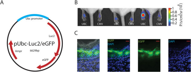
Figure 1. Detection of reporter plasmid expression in mouse footpad skin.

(A) Schematic map of pUbc-luc2/eGFP plasmid. (B) Bioluminescence imaging of pUbc-luc2/eGFP and pCMV-hMGFP/CBL (left and right paws, respectively) expression 24 h after intradermal paw injection. (C) Fluorescence microscopy of a skin section obtained from a paw previously imaged for bioluminescence (see Panel B, mouse 3, left paw) injected with pUbc-luc2/eGFP (scale bar is 50 µm). DAPI stain (blue) was used to identify cellular nuclei. Left panel shows brightfield overlay. Abbreviations: Ampr, ampicillin resistance; eGFP, enhanced green fluorescence protein; luc2, luciferase 2; Ubc, ubiquitin c; p, photons; sr, steradian.
