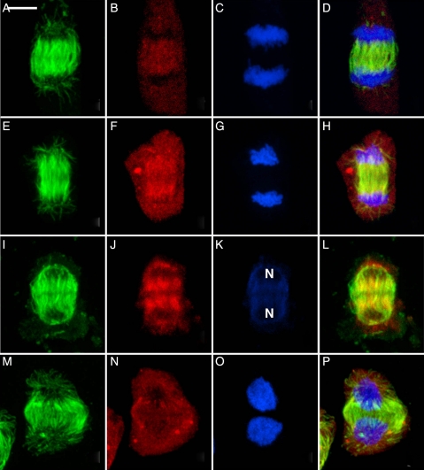
Fig. 5.
Telophase showing the localization of γ-tubulin at the proximal faces of the reforming nuclei where it is associated with development of the bipolar phragmoplast arrays. Scale bar = 5 µm. (A, E, I, M) Microtubules; (B, F, J, N) γ-tubulin; (C, G, K, O) nuclei; (D, H, L, P) composite. (A–D) The liverwort R. hemisphaerica. (E–H) The moss P. pyriforme. (I–L) The hornwort P. laevis. The daughter nuclei, weakly stained by To-Pro-3, are labelled (N). (M–P) The expanded phragmoplast in R. hemisphaerica.

