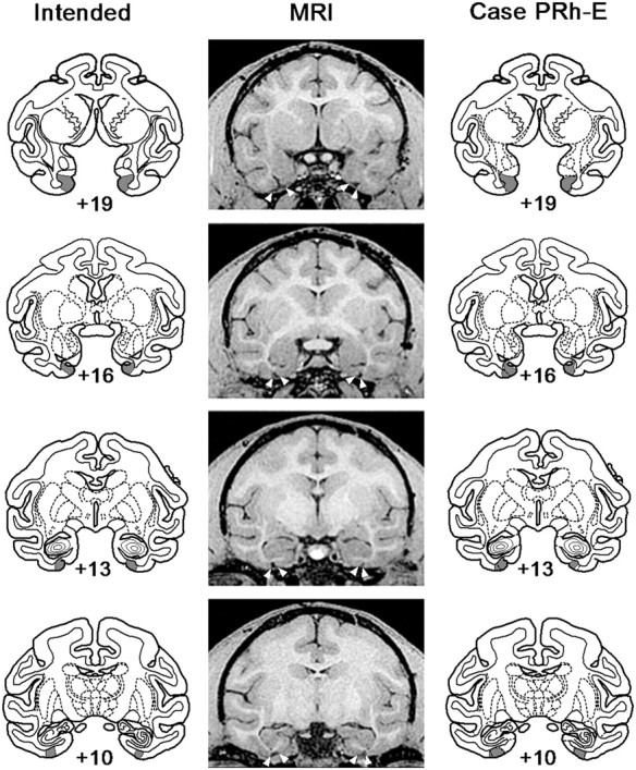
Figure 2.

Location and extent of the perirhinal cortex lesion in PRh-E. The intended lesion of perirhinal cortex (shaded region) is shown on coronal sections from a standard rhesus monkey brain (left column). Postoperative MR images from matching levels (middle column) and plots of the lesion (shaded region) onto sections (right column) show the extent of the lesion in PRh-E. Numerals indicate distance in millimeters from the interaural plane. White arrows in the MR images show the boundaries of the lesion.
