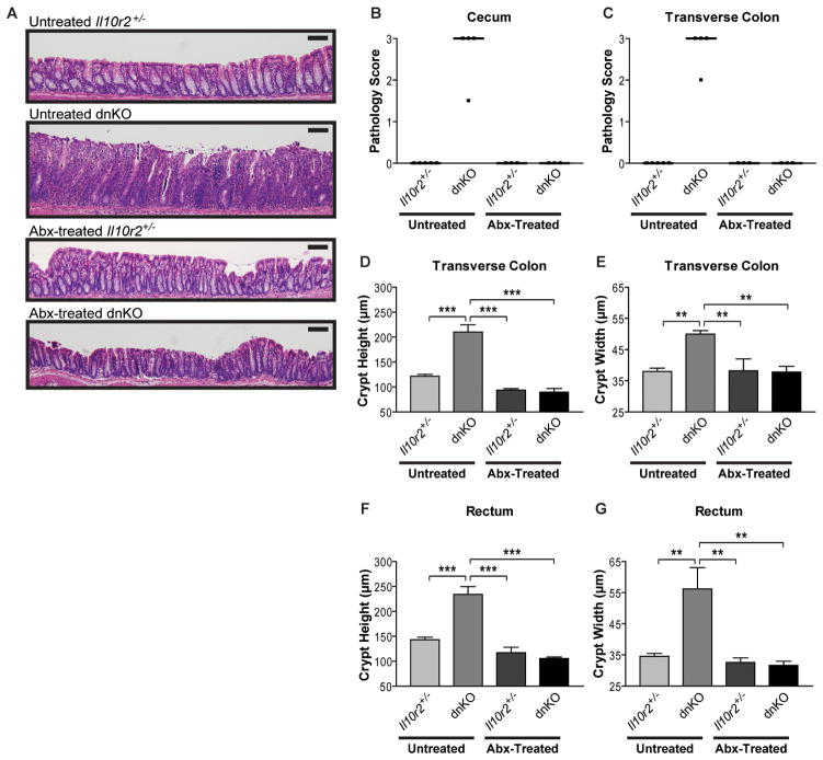
Figure 1. Antibiotic treatment quantitatively prevents colitis development in dnKO mice.
(A) Representative images of H&E stained rectal histology of 4-week-old untreated and antibiotic-treated Il10r2+/− and dnKO mice (distal 0.5 cm of colon). Abx = antibiotics (metronidazole + ciprofloxacin in drinking water). Scale bar = 100 μm.
(B and C) Cecum and transverse colon pathology scores of untreated and antibiotic-treated Il10r2+/− and dnKO mice as described in Figure 1A. Intestinal whole mounts were scored for gross pathology in a blinded fashion by an anatomic pathologist (T.S.S.) according to a validated system: ranging from 0 (no pathology) to 3 (severe pathology; see Figure S1 for details). Individual (squares) and median (bars) pathology scores are displayed. Kruskal-Wallis test: (B) H3 = 13.75, p = 0.0033; (C) H3 = 13.75, p = 0.0033. (D to G) Transverse colon and rectum crypt heights and crypt widths of mice in described in Figures 1B and 1C displayed as mean +/− SEM. 1-way ANOVA with post-hoc Tukey’s test: (D) F3,11 = 40.87, p < 0.0001; (E) F3,11 = 10.71, p = 0.0014; (F) F3,11 = 33.25, p < 0.0001; (G) F3,11 = 9.501, p = 0.0022. All statistically significant pairwise comparisons are displayed: **, p < 0.01, ***, p < 0.005.
See also Figure S1

